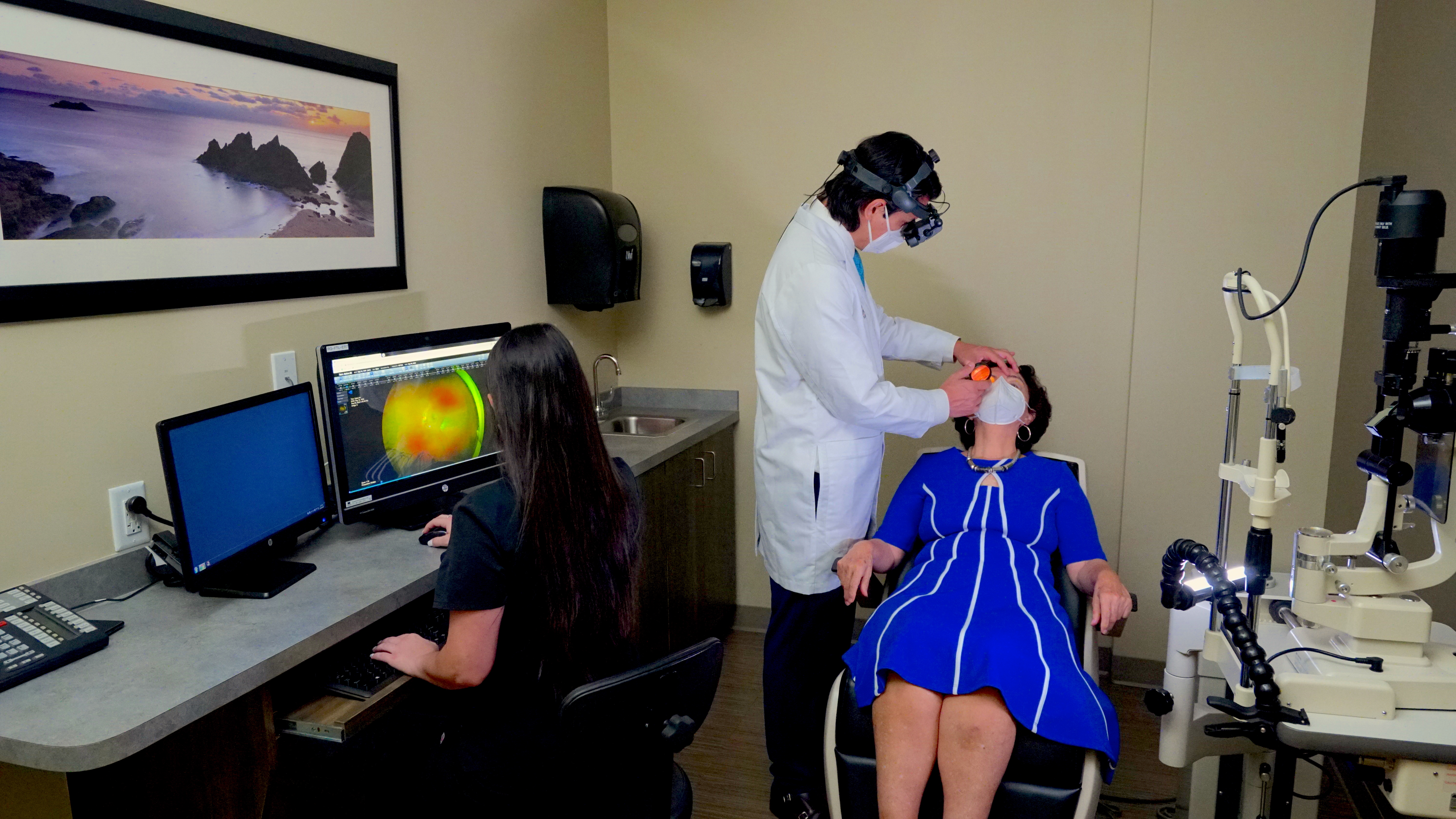
Retinal Diagnostic Procedures: What to Expect

Lining the back of the eye is a thin layer of light-sensitive tissue known as the retina. Measuring between 30 and 40 mm in diameter and 0.5 mm in thickness, this delicate tissue is the primary mechanism responsible for our ability to see. When the retina suffers damage from illness or injury, our vision and, by extension, our overall quality of life, can be significantly impacted.
Because of the retina’s location along the back of the eye, a retinal diagnostic exam requires sophisticated techniques and technologies to assess the presence and/or severity of retinal damage. In comparison to other types of eye exams, a retinal diagnostic exam tends to be very involved and can take more time to complete. Here’s what patients can expect to happen during a retinal diagnostic exam.
Dilated Eye Exam
The exam begins with a dilated eye exam, which involves applying special drops to the eyes that cause the pupil to widen. The pupil is the black circle that lies in the center of the iris that allows light to enter the eye and travel to the retina. The size of the pupil is governed by the muscular tissue of the iris, which controls the amount of light that enters the eye and reaches the retina. When we are in the presence of brighter light, our pupil typically contracts and gets smaller. In dim light or darkness, the pupil expands to allow more light into the eye.
The dilating eye drops stop the iris from contracting and making the pupil smaller, which makes it possible for retina specialists to examine the retina by shining a light into the eye. It also makes it possible for retina specialists to take digital images of the retina. Prior to dilating the pupils, the doctor will test for visual acuity to determine if the patient is experiencing any visual problems.
Retinal Imaging
A retinal exam usually consists of one or more imaging techniques. One of the most common imaging tests used by retina specialists is optical coherence tomography (OCT). This non-invasive imaging technique uses an array of light to scan the inside of the eye and capture high-definition images of the retinal tissue. It is used to detect and monitor damage in the retina and optic nerve.
Another common imaging technique is fluorescein angiography, which involves a specialized camera and a photosensitive dye called fluorescein. The dye is injected into the arm so that it can travel to the retinal vascular system, where it will highlight the blood vessels. Images are then captured to see if there are any abnormal blood vessels or retinal damage. This is particularly useful for diagnosing and monitoring conditions such as diabetic retinopathy, wet age-related macular degeneration, and retinal vein and artery occlusions.
After a Retinal Exam
It can take several hours for the effects of the dilating eye drops to wear off. During this time, your eyes will be more sensitive to light. Blurry vision is also common. It’s recommended that patients undergoing a retinal exam have someone with them who can drive them home afterward. While there are no side effects associated with OCT, there are some minor side effects of fluorescein angiography, including a temporary yellow discoloration of the skin and dark orange urine.
For advanced retinal diagnostic testing and treatment in South Florida, contact Retina Group of Florida today.


